AMS as a Tool in the Biological Sciences
Radioisotope labeling has been an important tool in the biological sciences and will continue to be used for many complex problems. Detection of radioisotopes is traditionally performed by decay counting, which is often limited by high backgrounds, as well as low specificity and low decay counting efficiency.
Mass spectrometry (MS) techniques, where individual nuclei are counted independent of decay, offers many advantages for long-lived radioisotope detection.
However, until the advent of AMS, measurement of 14C by MS has been fraught with difficulty, mostly due to problems in resolution and isobaric interference. Since the late 1980s, AMS has evolved as a biomedical tool, offering the required sensitivity, selectivity, and precision to address questions that alternative methodologies have been unable to achieve in practice. The sensitivity of AMS allows biological tracer studies to be conducted in humans with applications in metabolism, toxicology, personalized medicine, and drug development.
| Year | Event |
|---|---|
| 1987 | Conception of using AMS for low dose biological applications. |
| 1989 | First BioAMS experiments conducted. BioAMS patents conceived. |
| 1990 | Collaborative work with University of California (UC) begins. |
| 1993 | First BioAMS patents awarded to LLNL. |
| 1994 | First human subject BioAMS studies conducted. |
| 1995 | UC- LLNL Campus laboratory Collaboration for BioAMS established. |
| 1999 | NIH Funded National Center for BioAMS established. |
| 2001 | LLNL - UC Davis Cancer Center alliance established. |
| 2001 | Dedicated BioAMS spectrometer installed. |
| 2002 | BioAMS patents licensed. |
| 2004 | National Center grant for BioAMS renewed. |
| 2006 | Two licensees open commercial BioAMS facilities in US. |
| 2009 | National Center grant for BioAMS renewed for second time. |
| 2011 | Online liquid sample analysis capability established. |
| 2014 | Liquid sample AMS patent awarded to LLNL. |
| 2014 | National Center grant for BioAMS renewed for third time. |
| 2014 | 250KV single stage AMS deck installed in a biomedical laboratory. |
Our work has developed powerful diagnostic tools and answered fundamental biomedical questions. Two such examples are: Development of a B12 bioavailability assay and evidence of cardiomyocyte renewal in humans.
B12 Assay
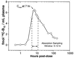
Assaying and understanding absorption and uptake of B12 is important because defects can lead to hematological and neurological complications. Accelerator mass spectrometry is uniquely suited for assessing absorption and kinetics in 14C-labeled substances after oral ingestion because it is more sensitive than decay counting and can measure levels of 14C in microliter volumes of biological samples with negligible exposure of subjects to radioactivity.
The assay employs amounts of B12 in the range of normal dietary intake. In a human kinetic study, a physiological dose (1.5 μg, 2.2 kBq/59 nCi) of purified 14C-B12 was administered and showed plasma appearance and clearance curves consistent with the predicted behavior of the pure vitamin. This method opens new avenues for study of B12 assimilation. This assay uses a micro-dose of 14C-labeled B12 (1/10th recommended daily allowance) and enables a simple test requiring one drop of blood to determine B12 absorption.
Proc. Natl. Acad. Sci, USA, February 22, 2006 103: 5694-5699. doi: 10.1073/pnas.0601251103
Cardiomyocyte Renewal
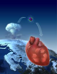
It has been difficult to establish whether we are limited to the heart muscle cells we are born with or if cardiomyocytes are generated also later in life. We have taken advantage of the integration of carbon-14, generated by nuclear bomb tests during the Cold War, into DNA to establish the age of cardiomyocytes in humans. We have found that cardiomyocytes renew, with a gradual decrease from 1% turning over annually at the age of 25 to 0.45% at the age of 75. Fewer than 50% of cardiomyocytes are exchanged during a normal life span. The capacity to generate cardiomyocytes in the adult human heart suggests that it may be rational to work toward the development of therapeutic strategies aimed at stimulating this process in cardiac pathologies.
"….this is one of those important studies that is going to change the way that we think for a very long time,"
Dr. Richard T. Lee, Brigham and Women's Hospital,
Boston, MA. CBS national evening news, April 3 2009.
Science 3 April 2009: 324: 98-102 doi: 10.1126/science.1164680
Today, nearly all our Biomedical AMS (BioAMS) work is conducted through our National Institutes of Health (NIH) funded National Resource for Biomedical Accelerator Mass Spectrometry and CAMS is the undisputed world leader in the application of AMS to biomedical science. This resource has been established to make AMS technology available to biomedical researchers who have a need for accurately measuring very low levels of 14C and 3H. This resource seeks to both develop the methods and instrumentation to make AMS a general use tool for biomedical researchers and to make it available for investigators needing access to techniques for the ultra-trace analysis of radioisotopes in biological studies.


